Microscope Worksheet Answers PDF: A Comprehensive Guide
Navigating the world of cellular biology often begins with mastering the microscope, and worksheets serve as crucial tools for reinforcing learning. These PDF resources
provide exercises on parts identification, magnification calculations, and observational drawing, aiding comprehension and skill development.
What are Microscope Worksheets?
Microscope worksheets are educational resources designed to enhance understanding of microscopy techniques and biological structures. Typically available as PDF documents, they present a variety of exercises focused on both theoretical knowledge and practical application. These worksheets commonly include tasks like identifying microscope parts – such as the ocular lens, objective lenses, stage, and light source – through labeling diagrams or multiple-choice questions.
Furthermore, they often require students to practice calculating magnification, a fundamental skill in microscopy. A significant component involves drawing and labeling specimens observed under different magnifications, fostering observational skills. Some worksheets delve into the cell cycle, mitosis, and even explore connections to concepts like cancer, utilizing interactive elements and answer keys for self-assessment. They are invaluable tools for reinforcing concepts learned in the lab or classroom.
Why Use Microscope Worksheets?
Microscope worksheets offer a structured approach to learning and solidifying microscopy skills. They bridge the gap between theoretical knowledge and practical application, allowing students to actively engage with the material. Utilizing these PDF resources reinforces understanding of microscope anatomy, magnification principles, and specimen observation techniques.
Worksheets provide a valuable self-assessment tool, with many including answer keys for immediate feedback. They help identify areas where students may need further clarification or practice. Moreover, they cultivate essential scientific skills like observation, drawing, and data interpretation. By consistently completing these exercises, students build confidence and competence in using a compound microscope, preparing them for more advanced biological studies and laboratory work. They are crucial for consolidating understanding of cell division and related concepts.
Types of Microscope Worksheets Available as PDFs
PDF worksheets encompass diverse exercises: identifying parts, mastering compound microscope usage, and calculating magnification—catering to varied learning needs and skill levels.
Identifying Microscope Parts Worksheets
These worksheets are foundational, focusing on recognizing the various components of a microscope. Students typically encounter exercises requiring them to label diagrams, often with multiple-choice options provided for each part. Common components featured include the arm, base, objective lenses, ocular lens (eyepiece), and stage.
Worksheets may present diagrams with numbered parts, demanding students to match the numbers with corresponding labels from a word bank; Alternatively, they might involve identifying parts directly from a microscope image. Some advanced worksheets challenge students to describe the function of each part, solidifying their understanding beyond simple recognition. These exercises are crucial for building a strong base for more complex microscopy tasks.
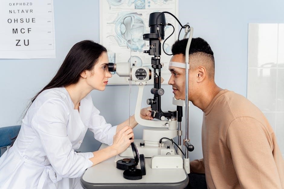
Using the Compound Microscope Worksheets
These worksheets bridge the gap between identifying parts and actually using a compound microscope. Exercises often involve step-by-step instructions for preparing a wet mount slide, focusing the microscope correctly, and adjusting illumination using the light source and diaphragm. Students may be asked to predict what they’ll observe at different magnification levels.
A common task is following procedures to view a specimen, then answering questions about their observations. Worksheets frequently include scenarios requiring students to troubleshoot focusing issues or optimize image clarity. Some exercises ask students to draw what they see at various magnifications, reinforcing observational skills. Mastering these practical skills is essential for successful microscopic investigation.
Calculating Magnification Worksheets
Magnification worksheets focus on the fundamental principle of total magnification – the product of the ocular lens magnification and the chosen objective lens magnification. Students practice applying the formula: Total Magnification = (Ocular Lens) x (Objective Lens). Problems range from simple calculations with provided lens powers to more complex scenarios requiring students to determine magnification given image size or vice versa.
These exercises often present different objective lenses (e.g., 4x, 10x, 40x, 100x) and ask students to calculate the resulting total magnification. Worksheets may also include questions about the relationship between magnification and field of view – understanding that as magnification increases, the field of view decreases. Accurate calculation skills are vital for interpreting microscopic observations.
Key Microscope Parts & Their Functions (Commonly Featured in Worksheets)
Worksheets consistently emphasize core components like the ocular lens, objective lenses, stage, light source, arm, and base, testing functional understanding.
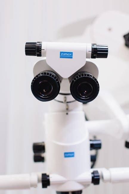
Ocular Lens (Eyepiece)
Microscope worksheets frequently assess understanding of the ocular lens, also known as the eyepiece. This is the lens through which the observer looks to view the specimen. Typically, it offers a magnification of 10x, although variations exist. Worksheets often ask students to identify the ocular lens on a diagram or describe its function in relation to total magnification.
A key concept tested is how the ocular lens’s magnification combines with the objective lens’s magnification to determine the overall magnification. For example, a 10x ocular lens paired with a 40x objective lens results in a total magnification of 400x. Questions may involve calculating total magnification given the individual lens powers. Furthermore, some worksheets explore the role of the eyepiece in forming a virtual, magnified image of the specimen.
Objective Lenses
Microscope worksheets dedicate significant attention to objective lenses, the primary lenses responsible for magnifying the specimen. These lenses typically range in magnification from 4x to 100x, and are crucial for initial image formation. Worksheets commonly require students to identify different objective lenses on microscope diagrams and state their corresponding magnification powers.
A frequent exercise involves calculating total magnification, demanding students multiply the objective lens magnification by the ocular lens magnification (usually 10x). Understanding the purpose of the oil immersion lens (100x) is also often tested, including when and why it’s used. Worksheets may present scenarios requiring students to select the appropriate objective lens for viewing specimens of varying sizes or detail. Correct identification and magnification calculations are key assessment points.
Stage & Stage Clips
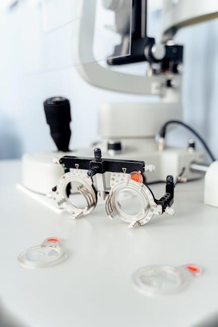
Microscope worksheets consistently feature questions regarding the stage and stage clips, fundamental components for specimen handling. The stage is the flat platform where slides are placed for observation, and worksheets often require students to identify its function in relation to focusing and viewing. Diagrams frequently ask for labeling of the stage’s mechanical components, like adjustment knobs for precise slide movement.
Stage clips secure the slide in place, preventing unwanted shifting during observation. Worksheets assess understanding of how these clips function and their importance for maintaining a stable image. Exercises may involve describing proper slide placement techniques or explaining the consequences of a poorly secured specimen. Comprehension of stage manipulation is vital for systematic specimen examination, a key skill reinforced by these educational resources.
Light Source & Diaphragm
Microscope worksheets frequently test understanding of the light source and diaphragm, critical for image clarity and contrast. The light source, traditionally a lamp, illuminates the specimen from below, and worksheets often ask students to explain its role in visualization. Questions may involve identifying the type of light source used or describing how its intensity affects the observed image.
The diaphragm controls the amount of light reaching the specimen, influencing both brightness and depth of field. Worksheets commonly present scenarios requiring students to predict how adjusting the diaphragm will impact image quality. Exercises might involve explaining the relationship between diaphragm aperture and resolution, or describing how to optimize light levels for different specimen types. Mastering these controls is essential for effective microscopic observation.

Arm & Base
Microscope worksheets consistently assess knowledge of the arm and base, fundamental components for support and transport. The arm serves as the primary handling point, connecting the body tube to the base, and worksheets often feature diagrams requiring students to correctly identify it. Questions may focus on proper carrying techniques, emphasizing the importance of using both hands – one on the arm and one under the base.
The base provides stable support for the entire microscope. Worksheets frequently include scenarios testing understanding of balance and safe handling. Exercises might involve explaining why a secure base is crucial for obtaining clear images or describing potential consequences of an unstable setup. Recognizing these structural elements is vital for responsible microscope operation and longevity.

Common Questions on Microscope Worksheets & Their Answers
Worksheets commonly pose questions about magnification, parts identification (often multiple choice), and specimen drawing/labeling, testing core microscopy skills and knowledge retention.
Magnification Calculation Questions
Magnification problems are a staple of microscope worksheets, designed to assess understanding of total magnification. Students frequently encounter questions requiring them to calculate total magnification by multiplying the ocular lens magnification (typically 10x) by the objective lens magnification (e.g., 4x, 10x, 40x, 100x).
Worksheets often present scenarios where students are given the magnification of both lenses and asked to determine the total magnification. Conversely, some questions might provide the total magnification and one lens’s magnification, requiring students to solve for the unknown magnification.
More complex problems may involve calculating the actual size of a specimen, utilizing magnification and a known scale. Understanding these calculations is fundamental to interpreting microscopic observations accurately. Correct answers demonstrate a grasp of the relationship between lens powers and the resulting image size.
Parts Identification Questions (Multiple Choice)
Multiple-choice questions focusing on microscope parts are incredibly common within these worksheets. Students are typically presented with a diagram of a microscope and asked to identify specific components from a list of options. These questions test recognition of key structures like the objective lenses, ocular lens (eyepiece), stage, stage clips, arm, base, light source, and diaphragm.
The questions often require differentiating between similar parts or understanding the function associated with each component. For example, a question might ask, “Which part controls the amount of light passing through the specimen?” with options like condenser, objective, or ocular lens.
Successfully answering these questions demonstrates a foundational understanding of microscope anatomy, crucial for proper usage and observation.
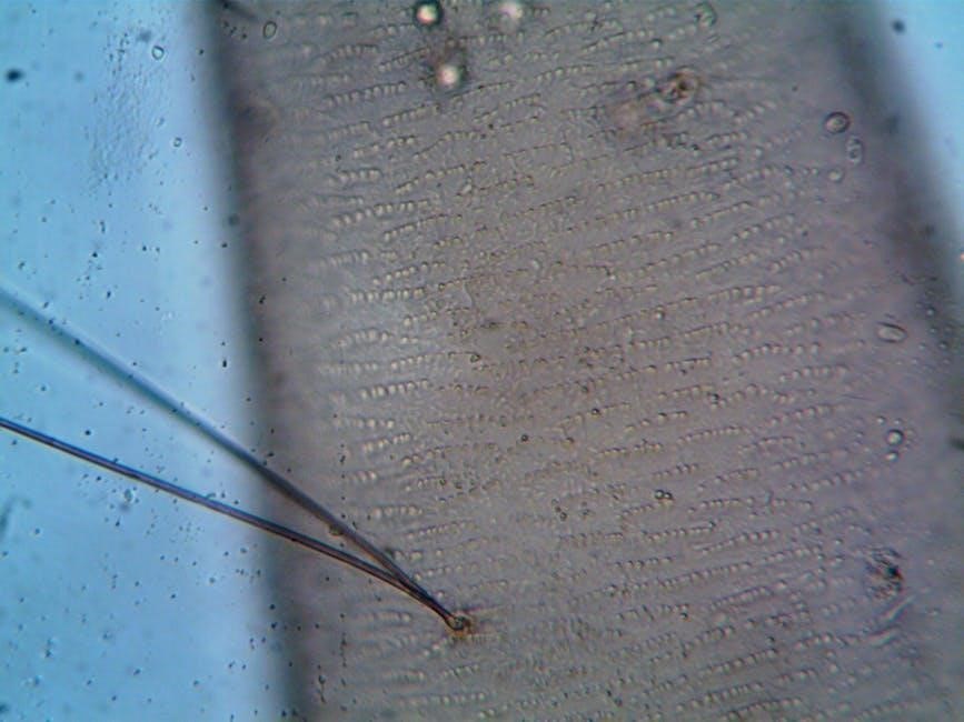
Drawing & Labeling Specimen Questions
A significant portion of microscope worksheets involve drawing and labeling specimens observed under different magnifications. Students are presented with a microscopic view – often a simple cell structure – and tasked with accurately recreating it on paper. This isn’t merely about artistic skill; it’s about developing observational abilities and understanding scale.
Worksheets frequently require students to label key features of the specimen, demonstrating their knowledge of cellular components. They may also need to indicate the magnification used for the drawing. This reinforces the relationship between magnification power and the apparent size of objects.
These exercises cultivate precision and attention to detail, essential skills for scientific illustration and data recording.

Where to Find Microscope Worksheet PDFs
Numerous online platforms and educational resources offer free microscope worksheets in PDF format, including science teacher websites and broader online learning hubs.
Educational Websites & Resources
A wealth of educational websites dedicate sections to science education, frequently including downloadable microscope worksheets as PDF files. These resources often cater to various grade levels, offering worksheets ranging from basic parts identification for elementary students to more complex magnification calculations for high school learners.
Many sites provide answer keys alongside the worksheets, facilitating self-assessment and independent study. Look for resources from established educational organizations or university science departments for reliable and accurate content. Some websites offer interactive worksheets that can be completed online, providing immediate feedback. Exploring these platforms can significantly enhance understanding of microscope techniques and biological concepts. Remember to check the website’s terms of use regarding downloading and distributing the materials.
Science Teacher Resources
Dedicated online platforms cater specifically to science educators, offering curated collections of microscope worksheets in PDF format. These resources are often designed by experienced teachers and align with national science education standards. Websites like Teachers Pay Teachers and science-focused educational marketplaces host a diverse range of worksheets, including those focusing on parts labeling, magnification exercises, and specimen drawing.
Many teacher resources include detailed answer keys and accompanying lesson plans, streamlining classroom preparation. Some platforms offer customizable worksheets, allowing teachers to tailor the content to their students’ specific needs. Utilizing these resources can save valuable time and ensure that students receive high-quality, standards-aligned instruction on microscope use and biological observation.
Online Learning Platforms
Increasingly, interactive online learning platforms are integrating microscope worksheets into their science curricula, often providing them as downloadable PDFs. Platforms like Khan Academy and Coursera, while not exclusively focused on worksheets, may offer related exercises as part of broader biology modules. These platforms frequently incorporate self-grading features and immediate feedback, enhancing the learning experience.
Furthermore, specialized educational websites dedicated to virtual labs and science simulations often include printable worksheets to complement their interactive content. These resources can be particularly valuable for remote learning or for students needing extra practice. Accessing these platforms often requires a subscription or enrollment in a course, but the benefits of structured learning and readily available resources can be substantial.

Tips for Completing Microscope Worksheets
Success with these PDFs hinges on solid microscope anatomy knowledge, magnification understanding, and diligent practice sketching observations accurately – key skills for biology!
Review Microscope Anatomy
Before tackling any worksheet, a thorough review of microscope parts is essential. Many worksheets focus heavily on identifying components like the ocular lens (eyepiece), objective lenses, stage, stage clips, light source, diaphragm, arm, and base. Familiarize yourself with the function of each part – how does the diaphragm control light, and what’s the purpose of the stage clips?
Worksheets often present diagrams requiring labeling, or multiple-choice questions testing your recognition. Understanding where each part is located and its specific role will dramatically improve your accuracy. Don’t just memorize names; visualize how they work together to create a magnified image. Resources detailing microscope anatomy are readily available online and in textbooks, providing valuable support for worksheet completion and overall comprehension.
Understand Magnification Principles

Mastering magnification calculations is a cornerstone of microscope worksheet success. Worksheets frequently present problems requiring you to determine total magnification by multiplying the ocular lens magnification by the objective lens magnification. For example, a 10x ocular lens combined with a 40x objective lens yields a total magnification of 400x.
Beyond simple calculation, grasp the concept of how magnification affects the field of view – higher magnification means a smaller field of view. Practice converting between magnification and field of view size. Understanding these principles isn’t just about getting the right answer on a worksheet; it’s about interpreting the scale of what you’re observing under the microscope and accurately documenting your findings.
Practice Drawing Observations
Microscope worksheets often require students to draw what they observe through the eyepiece, a skill vital for scientific documentation. Focus on accurately representing shapes, sizes, and relative positions of cellular structures. Don’t aim for artistic perfection; prioritize clarity and detail. Labeling is crucial – clearly identify key components of the specimen you’ve drawn.
Consistent practice improves observational skills and reinforces understanding of microscopic anatomy. Start with simple specimens and gradually increase complexity. Pay attention to magnification; your drawing should reflect the scale at which you viewed the sample. Remember, a well-executed drawing is a powerful tool for communicating scientific observations and solidifying your learning.